
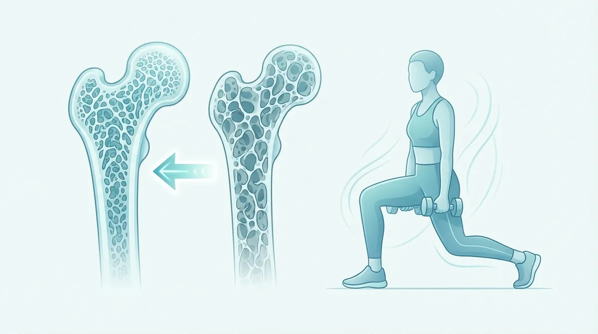
Title: Osteoporosis and Physical Exercise: Weight-Bearing Exercises to Strengthen Bones
The aging of the skeletal system and the consequent loss of bone mineral density represent one of the most relevant clinical challenges in today’s rehabilitative and preventive medicine. Optimal management of this condition requires a multidisciplinary approach, within which the combination of osteoporosis and physical exercise constitutes one of the fundamental pillars for the preservation of bone microarchitecture and the prevention of fragility fractures. Osteoporosis is a systemic skeletal disease characterized by a reduction in bone mass and a deterioration of bone tissue structure, factors that lead to increased fragility and the consequent risk of fractures, particularly affecting the femur, vertebrae, and wrist. Although pharmacological and nutritional intervention is often necessary, international scientific literature agrees that mechanical stimulation induced by movement is essential for maintaining skeletal homeostasis. The aim of this article is to analyze in a detailed and scientific manner the physiological mechanisms, international guidelines, training protocols, and necessary precautions for structuring an effective and safe exercise program, particularly aimed at the over 50 population. It is fundamental to state that any rehabilitative or training intervention must always be preceded by a careful clinical evaluation; therefore, before undertaking any new exercise program, it is imperative to consult your doctor or physical therapist for an accurate diagnostic assessment.
- Osteoporosis involves bone loss and structural deterioration, significantly increasing fracture risk.
- Physical exercise is crucial for preserving bone microarchitecture and preventing fragility fractures.
- Optimal osteoporosis management requires a multidisciplinary approach, integrating exercise.
- Exercise interventions for osteoporosis require careful planning and medical supervision.
The Link Between Osteoporosis and Physical Exercise: Physiology of Bone Remodeling
To fully understand the effectiveness of osteoporosis and physical exercise, it is necessary to explore the complex physiological mechanisms that govern bone remodeling. Bone tissue is not an inert structure, but rather a dynamically active organ, in continuous renewal through a coordinated process of resorption and neoformation. This process is orchestrated by two main cell types: osteoclasts, responsible for the breakdown of old or damaged bone matrix, and osteoblasts, responsible for the synthesis of new osteoid matrix and its subsequent mineralization.
The fundamental biomechanical principle linking mechanical load to bone density is known as Wolff’s Law (Wolff, 1892), which postulates that bone adapts to the loads placed upon it. If the load increases, the bone remodels to become stronger and resist such stress; conversely, in the absence of load (as occurs in prolonged immobilization or in zero gravity), bone mass rapidly decreases. At the cellular level, this phenomenon is mediated by mechanotransduction. Osteocytes, cells trapped within the mineralized bone matrix, act as true mechanoreceptors. When bone undergoes elastic deformation (strain) due to mechanical load, fluid flows are generated within the bone canaliculi. Osteocytes perceive this fluid shear stress and, in response, release biochemical signals, such as nitric oxide and prostaglandins, which inhibit osteoclast activity and stimulate osteoblast proliferation and activity (Rubin et al., 2001).
However, not all mechanical stimuli are equally effective. The “Mechanostat” theory (Frost, 2003) suggests that there is a minimum threshold of bone deformation (measured in microstrain) necessary to trigger the osteogenic response. Static or repetitive low-intensity stimuli tend to rapidly saturate mechanoreceptors, making the bone refractory to further stimuli. Conversely, dynamic loads of unusual magnitude, applied with a high strain rate and interspersed with rest periods, prove to be the most powerful inducers of bone formation. This explains why specific types of training are significantly superior to others in counteracting skeletal demineralization.
Epidemiological Data and Occupational Impact: INAIL Statistics
The impact of osteoporosis is not limited to the personal clinical sphere, but extends significantly to the occupational and socio-economic context. The progressive aging of the workforce has made musculoskeletal pathologies, and osteoporosis in particular, a central theme for occupational medicine and safety in professional environments. According to data and publications from the National Institute for Insurance against Accidents at Work (INAIL), female and male workers over 50 represent a particularly vulnerable population group to the consequences of bone fragility.
INAIL statistics highlight how fragility fractures (which occur due to low-energy trauma that would not cause injury in a healthy bone) are a primary cause of prolonged absenteeism, temporary or permanent disability, and, in many cases, unsuitability for specific job tasks. The professional sectors most at risk are those that require manual handling of loads, prolonged incongruous postures, or that present a high risk of slipping and falling. These include the healthcare and social assistance sector (nurses, healthcare support workers), the industrial cleaning sector, logistics, and agriculture.
INAIL emphasizes the importance of primary and secondary prevention in the workplace. For workers diagnosed with osteopenia or osteoporosis, the risk of vertebral compression fractures during lifting loads, even of modest entity, increases exponentially. Therefore, health surveillance must include a careful assessment of biomechanical risk. The implementation of corporate preventive exercise programs and ergonomics education are strongly recommended strategies. In this context, targeted physical activity not only improves bone density but also increases muscle strength and balance, drastically reducing the incidence of work-related injuries due to accidental falls. It is always advisable for the worker to report their condition to the occupational physician and rely on a doctor or physical therapist for the development of a functional recovery plan compatible with their job duties.
International Guidelines for Osteoporosis: Physical Exercise and Types of Load
The prescription of osteoporosis physical exercise should be considered akin to a pharmacological prescription, with precise indications on dosage, frequency, intensity, and type. Major international scientific societies, including the American College of Sports Medicine (ACSM) and the National Osteoporosis Foundation (NOF), have drawn up detailed guidelines to maximize osteogenic benefits while minimizing the risk of injury. Exercise interventions are divided into three main macro-categories: weight-bearing aerobic exercises, resistance and muscle strength training, and impact exercises.
Weight-Bearing Aerobic Exercises
Weight-bearing aerobic exercises are all those activities in which the body’s weight is supported by the lower limbs, forcing the skeleton to work against the force of gravity. Brisk walking, jogging, climbing stairs, dancing, and hiking fall into this category. These activities are fundamental for maintaining bone mineral density (BMD) at the femoral neck and lumbar spine, the sites most exposed to fracture risk.
It is of crucial importance to distinguish these activities from non-weight-bearing ones. Swimming and cycling, while excellent for cardiovascular health and muscle conditioning without joint stress, do not provide the mechanical stimulus necessary to induce osteogenesis (Gomez-Cabrera et al., 2008). Water cancels the effect of gravity, and the bicycle supports the body’s weight on the saddle. Therefore, although these activities can be part of a general wellness program, they cannot constitute the primary intervention for the treatment of osteoporosis.
Resistance and Muscle Strength Training (Resistance Training)
Resistance training, which involves the use of free weights, isotonic machines, elastic bands, or one’s own body weight (calisthenics), is considered the most effective intervention for increasing bone and muscle mass. Intense muscle contraction exerts direct traction on tendon insertions at the periosteum, generating local deformation forces (strain) much greater than those produced by simple gravity.
Scientific literature (Daly et al., 2019) demonstrates that high-intensity resistance training (HIRT) can produce significant improvements in BMD even in postmenopausal women with established osteoporosis, provided it is performed under strict supervision. Exercises should be multi-joint and involve large muscle groups. Classic examples include squats, lunges, deadlifts (performed with impeccable technique to protect the spine), overhead presses, and rows. Strengthening the back extensor muscles is particularly critical: strong paravertebral muscles reduce compressive load on the anterior vertebral bodies, decreasing the risk of hyperkyphosis and wedge fractures.
Impact Exercises
Impact exercises generate high and sudden ground reaction forces (GRF), which are transmitted through the skeleton. These forces create that high “strain rate” which, as postulated by the Mechanostat theory, is the most powerful stimulus for osteogenesis. Jumping rope, performing jumps (plyometrics), hopping in place, or vigorous step-ups are examples of high-impact exercises.
However, the administration of impact exercises requires extreme caution and risk stratification. In subjects with osteopenia or with healthy bones for preventive purposes, high impact is strongly recommended. Conversely, in subjects with severe osteoporosis (T-score less than -2.5) or with previous fragility fractures, high impact is contraindicated due to the high risk of causing acute injuries. In these cases, low impact is opted for, such as accentuated marching in place or the “heel drop” (rising onto the balls of the feet and letting the heels drop to the ground with controlled force), always with prior authorization from your doctor or physical therapist.
Specific Training Protocols for Prevention and Treatment
To translate physiological principles into clinical practice, it is necessary to structure training protocols following the FITT principle (Frequency, Intensity, Time, Type). A well-balanced program must integrate different exercise modalities to achieve a synergistic effect on bones, muscles, and the central nervous system.
Below is a summary table of the optimal parameters for structuring a training program aimed at bone health, based on current scientific evidence.
- Set elastici resistenza (5 livelli) (Esercizi | 12-25€)
- Foam roller alta densità (Auto-trattamento | 18-35€)
- Tappetino fitness antiscivolo (Esercizi | 20-40€)
| Type of Exercise | Frequency | Intensity | Time (Duration) | Practical Examples |
|---|---|---|---|---|
| Weight-Bearing Aerobic | 3-5 days a week | From moderate (able to talk with slight breathlessness) to vigorous. | 30-45 minutes per session (also broken into 10 min blocks). | Brisk walking, Nordic Walking, stair climbing, dancing. |
| Strength and Muscle Resistance | 2-3 days a week (non-consecutive) | High: 70-85% of 1RM (Repetition Maximum). 2-3 sets of 8-12 repetitions. | 20-30 minutes. Focus on large muscle groups. | Squats, leg press, pull-ups, rows, back extensions. |
| Impact (if tolerated) | 3-5 days a week | Variable based on BMD. 2 to 4 times body weight (GRF). | Short sessions: 50-100 total impacts per session, divided into sets. | Heel drops, hopping in place, dynamic step-ups. |
| Balance and Proprioception | Daily or almost daily (4-7 days) | Progressively challenging (reduced base of support, eyes closed). | 10-15 minutes per day. | Tai Chi, single-leg stands, tandem walking. |
Load Progression
An essential element for long-term success is load progression (Progressive Overload). Since bone adapts to mechanical stimulus, maintaining the same weight or routine for months will lead to a plateau, where bone remodeling will cease. It is necessary to gradually increase resistance (weights lifted), the complexity of exercises, or the magnitude of impact. This progression must be slow, constant, and continuously monitored. An increase of 5-10% of the load every 2-4 weeks is generally considered safe. However, execution technique must never be sacrificed to lift heavier loads. Initial supervision by a movement professional is essential to learn correct movement patterns. The need to consult your doctor or physical therapist to establish the most suitable progression parameters for the individual clinical picture is reiterated.
Contraindicated Exercises and Fundamental Precautions in Clinical Practice
While physical exercise is a powerful therapy, performing biomechanically incorrect movements can become a primary risk factor for fractures, especially at the spinal level. Vertebral bodies affected by osteoporosis lose their complex internal trabecular structure, becoming like “empty boxes” incapable of resisting asymmetric compression forces.
Movements to Absolutely Avoid
Biomechanics literature (Sinaki et al., 1984) has widely demonstrated that anterior spinal flexion, especially when associated with a load, dramatically increases pressure on the anterior portion of the thoracic and lumbar vertebral bodies. This mechanism is the main cause of wedge fractures (anterior compression fractures), which lead to progressive dorsal hyperkyphosis (the so-called “dowager’s hump”).
Therefore, the following movements and exercises are to be considered contraindicated in subjects with osteoporosis:
- Dynamic spinal flexion: Traditional abdominal exercises such as sit-ups, crunches, or machines that induce trunk flexion.
- Prolonged static spinal flexion: Touching toes with straight legs (toe touches), deep flexion stretching, yoga poses that require marked rounding of the back (e.g., plow pose).
- Spinal twisting under load: Movements that combine trunk flexion and rotation (e.g., shoveling, torso twisting machines in the gym, uncontrolled golf swings). The shear forces generated by twisting are extremely damaging to the annulus fibrosus of the intervertebral discs and to osteoporotic facet joints.
- Incorrect weight lifting: Picking up objects from the ground by bending the back instead of bending the knees and hips while keeping the spine in a neutral position (hip hinge).
Fall Risk Assessment and Environmental Safety
In addition to exercise biomechanics, the safety of the training environment is paramount. Femoral neck and wrist fractures (Colles’ fracture) occur almost exclusively as a result of a fall. Therefore, fall prevention is as important as increasing bone mass. During training, it is necessary to ensure that the environment is free of obstacles, slippery rugs, or poor lighting. The use of appropriate footwear, with non-slip soles and good arch support, is mandatory. In cases of known postural instability, standing exercises should be performed near a stable support (such as a heavy chair, a wall bar, or a handrail) to hold onto in case of loss of balance. Any training program must be approved by your doctor or physical therapist, who will pre-evaluate the patient’s fall risk through specific clinical tests (e.g., Timed Up and Go test, Berg Balance Scale).
The Role of Proprioception and Balance in Fracture Prevention
As mentioned, bone density is only one part of the equation in osteoporosis management; the other half is represented by trauma prevention. With advancing age, there is a physiological decline in sensory systems (visual, vestibular, somatosensory) and neuromuscular reaction times, factors that exponentially increase the risk of accidental falls. The integration of specific balance and proprioception training is therefore an indispensable requirement in any therapeutic protocol.
Proprioceptive training aims to improve body position awareness in space and refine automatic postural responses. Practices such as Tai Chi have been shown, in numerous randomized clinical trials, to significantly reduce the incidence of falls in the elderly, thanks to slow, controlled movements and continuous weight transfers from one limb to another. Other fundamental exercises include maintaining a standing position on one leg (single-leg stance), tandem walking (one foot directly in front of the other, as on a tightrope), and the use of unstable surfaces (such as proprioceptive cushions or wobble boards), although the latter should only be introduced in advanced stages and under strict supervision.
The goal of these exercises is to train balance recovery strategies (ankle strategy, hip strategy, and stepping strategy). A reactive neuromuscular system is able to correct a stumble before it turns into a disastrous fall, thus protecting the fragile skeleton from high-energy impacts that would inevitably lead to a fracture.
Nutrition and Lifestyle in Synergy with Training
Although the focus of this article is on physical exercise, it is important to emphasize that mechanical stimulation alone cannot build new bone matrix in the absence of the necessary biological “building blocks”. Training must be supported by adequate nutritional intake and a healthy lifestyle to maximize results.
Calcium and vitamin D are the key micronutrients for skeletal health. Calcium provides the mineral for bone rigidity, while vitamin D is essential for the intestinal absorption of calcium itself. Guidelines recommend an adequate calcium intake through diet (dairy products, dark green leafy vegetables, calcium-rich waters) and, if necessary, through supplementation. Similarly, safe sun exposure and potential vitamin D supplementation must be clinically evaluated. Furthermore, adequate protein intake is essential not only for bone matrix (which is largely composed of collagen) but also to support muscle hypertrophy induced by strength training.
Finally, it is imperative to eliminate modifiable risk factors that accelerate bone resorption. Cigarette smoking has a direct toxic effect on osteoblasts and alters estrogen metabolism, while alcohol abuse interferes with calcium absorption and increases the risk of falls due to impaired balance. The synergy between a well-structured exercise program, targeted nutrition, and the abandonment of unhealthy habits represents the most powerful conservative approach against the progression of osteoporosis.
Frequently Asked Questions (FAQ) about Osteoporosis and Physical Exercise
1. Can I lift weights if I have been diagnosed with osteoporosis?
Yes, weightlifting (resistance training) is not only permitted but strongly recommended by international guidelines. The use of overload stimulates the formation of new bone tissue and strengthens muscles, protecting joints. However, it is crucial that loads are introduced gradually, that the execution technique is perfect, and that spinal flexion and twisting movements are avoided. It is imperative to begin this journey under the supervision of your doctor or physical therapist to avoid injuries.
2. Is walking every day enough to treat osteoporosis?
No, walking alone is not enough. Although walking is an excellent weight-bearing aerobic activity that helps maintain femoral bone density and is good for the cardiovascular system, it does not provide a mechanical stimulus intense enough (high “strain”) to induce a significant increase in bone mass in the rest of the body, particularly in the spine and upper limbs. To achieve optimal results, walking must be supplemented with muscle strength and balance exercises.
3. Are swimming and aqua aerobics good for fragile bones?
Swimming and water activities are excellent for joint health (e.g., in cases of severe osteoarthritis), for the heart, and for general muscle tone, but they are not effective in stimulating bone mineral density. Water cancels the effect of gravity, eliminating the mechanical load necessary to activate osteoblasts (the cells that build bone). Therefore, those suffering from osteoporosis should prioritize “dry land” activities where the body must support its own weight against gravity.
4. What movements should I absolutely avoid in daily life and in the gym?
The most dangerous movements for those with osteoporosis are anterior spinal flexion (bending forward by rounding the back, such as to touch your toes or perform classic “crunch” abdominal exercises) and trunk twisting, especially if performed simultaneously or under
Frequently Asked Questions
What is osteoporosis?
Osteoporosis is a systemic skeletal disease characterized by a reduction in bone mass and a deterioration of bone tissue structure. These factors lead to increased bone fragility and a higher risk of fractures, particularly affecting the femur, vertebrae, and wrist.
Why is physical exercise considered a fundamental pillar in osteoporosis management?
Physical exercise provides essential mechanical stimulation that is crucial for maintaining skeletal homeostasis. This stimulation helps preserve bone microarchitecture and plays a vital role in preventing fragility fractures.
What type of exercise is generally recommended for strengthening bones in individuals with osteoporosis?
Weight-bearing exercises are particularly emphasized for their role in strengthening bones. These exercises induce mechanical stimulation, which is essential for maintaining bone mineral density and improving bone structure.
What is the importance of consulting a healthcare professional before starting an exercise program for osteoporosis?
A careful clinical evaluation by a doctor or physical therapist is imperative before undertaking any new exercise program. This ensures an accurate diagnostic assessment and allows for the structuring of an effective and safe intervention tailored to individual needs.
Sources and Scientific References
- Watson SL et al. (2018). High-Intensity Resistance and Impact Training Improves Bone Mineral Density and Physical Function in Postmenopausal Women With Osteopenia and Osteoporosis: The LIFTMOR Randomized Controlled Trial. J Bone Miner Res. 33:211-220. DOI | PubMed
- Linhares DG et al. (2022). Effects of Multicomponent Exercise Training on the Health of Older Women with Osteoporosis: A Systematic Review and Meta-Analysis. Int J Environ Res Public Health. 19. DOI | PubMed
- Morin SN et al. (2025). Osteoporosis: A Review. JAMA. 334:894-907. DOI | PubMed
- Beck BR et al. (2017). Exercise and Sports Science Australia (ESSA) position statement on exercise prescription for the prevention and management of osteoporosis. J Sci Med Sport. 20:438-445. DOI | PubMed
- Stanghelle B et al. (2020). Physical fitness in older women with osteoporosis and vertebral fracture after a resistance and balance exercise programme: 3-month post-intervention follow-up of a randomised controlled trial. BMC Musculoskelet Disord. 21:471. DOI | PubMed