
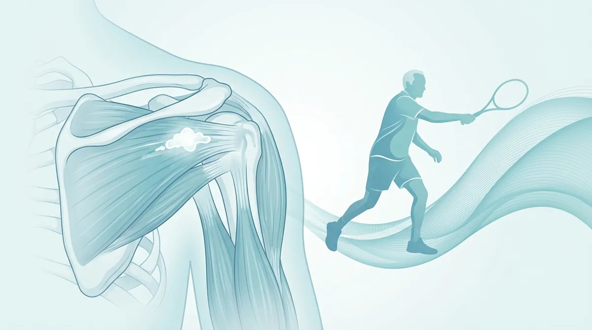
Title: Calcific Shoulder and Sport: How to Manage Calcific Tendinitis Over 50
Frequently Asked Questions
What is calcific tendinitis of the shoulder?
Calcific tendinitis is a condition characterized by the deposition of calcium hydroxyapatite crystals within the tendons of the rotator cuff, most commonly the supraspinatus muscle tendon. This can lead to pain, inflammation, and functional limitation in the shoulder.
- Calcific tendinitis involves calcium crystal deposition in rotator cuff tendons, causing pain.
- Athletes over 50 are susceptible due to aging tissues and biomechanical stress.
- It is a self-limiting condition with a cyclical evolution and resorptive phase.
- Symptoms range from asymptomatic to severe, often nocturnal, debilitating pain.
Why are athletes over 50 particularly susceptible to calcific tendinitis?
Athletes over 50 are more susceptible due to a combination of physiological tissue alterations related to aging and the ongoing biomechanical demands of their physical activity. Factors like tissue hypoxia and repeated microtrauma from prolonged biomechanical overload contribute to its development.
Is calcific tendinitis a permanent condition?
No, calcific tendinitis is not considered an irreversible degenerative process; it is a self-limiting pathology with a cyclical evolution. It progresses through distinct phases, including a resorptive phase where the body actively attempts to reabsorb the calcium deposit.
What are the typical symptoms of calcific tendinitis?
Symptoms can vary greatly, from being completely asymptomatic in the initial pre-calcific phase to experiencing dull discomfort during arm elevation. The most painful phase, known as the resorptive phase, can cause excruciating, often nocturnal, and debilitating pain due to a significant inflammatory response.
What should I do if I suspect I have calcific tendinitis?
If you suspect you have calcific tendinitis, it is crucial to seek evaluation and supervision from a doctor or physical therapist. Self-diagnosing or attempting DIY rehabilitation protocols could potentially worsen your clinical picture and delay appropriate treatment.
The management of tendinous pathologies represents a complex clinical challenge, especially when dealing with the delicate combination of calcific shoulder and sport in athletes over 50. Calcific tendinitis of the rotator cuff is a pathological condition characterized by the deposition of calcium hydroxyapatite crystals within the tendinous tissue, predominantly affecting the supraspinatus muscle tendon. In athletes over fifty years of age, this condition takes on particular contours due to the physiological tissue alterations related to aging, combined with the biomechanical demands of physical activity. The approach to this pathology requires a deep understanding of its etiopathogenesis, joint biomechanics, and the most advanced therapeutic strategies, in order to ensure a safe and high-performing return to sports practice. It is fundamental to emphasize that every diagnostic and therapeutic pathway must always be evaluated and supervised by a doctor or physical therapist, being wary of self-diagnoses and DIY rehabilitation protocols that could worsen the clinical picture.
The prevalence of calcific tendinitis in the general population varies from 2.7% to 20%, with a peak incidence precisely in the fifth and sixth decades of life (ElShewy et al., 2016). In master athletes, particularly those who practice “overhead” sports (such as tennis, swimming, volleyball, or golf), the presence of calcifications can significantly alter scapulohumeral kinematics, generating secondary subacromial impingement, acute pain, and functional limitation. This article exhaustively analyzes the nature of the pathology, risk factors, diagnostic methods, and the best scientific evidence related to conservative and rehabilitative treatment.
Calcific shoulder and sport: etiopathogenesis and phases of the disease
To fully understand the implications of **calcific shoulder and sport**, it is necessary to analyze the biological mechanisms that lead to the formation of calcium deposits. Contrary to what one might think, calcific tendinitis is not an irreversible degenerative process, but a self-limiting pathology characterized by a well-defined cyclical evolution. The most scientifically accredited theory is that proposed by Uhthoff (Uhthoff et al., 1997), which describes the pathology as a cell-mediated process, divided into three distinct phases.
Pre-calcific phase
In this initial phase, usually asymptomatic, fibrocartilaginous metaplasia of the tendinous tissue occurs. Tendon cells (tenocytes) transform into chondrocytes, creating an environment favorable for subsequent calcium deposition. This process appears to be triggered by a combination of tissue hypoxia (often localized in Codman’s “critical zone” of the supraspinatus tendon) and repeated microtrauma, factors frequently found in athletes over 50 due to prolonged biomechanical overload over the years.
Calcific phase
The calcific phase is in turn divided into three sub-phases:
- Formative phase: Calcium hydroxyapatite crystals are deposited in the extracellular matrix. The deposit has a chalky, hard consistency. In this phase, pain may be absent or present as a dull discomfort during arm elevation movements.
- Resting phase: The calcification is stable. The deposit is surrounded by a fibrous rim. Symptoms are generally mild, mainly related to the mechanical bulk of the deposit in the subacromial space during sports activity.
- Resorptive phase: This is the most painful and acute phase of the pathology. The immune system recognizes the calcium deposit as a foreign body and triggers a massive inflammatory response mediated by macrophages and multinucleated giant cells. The deposit takes on a toothpaste-like (pultaceous) consistency, and intratendinous pressure increases drastically, causing excruciating pain, often nocturnal and debilitating, which prevents any sports activity and even normal daily activities.
Post-calcific phase
Once the calcium deposit has been reabsorbed, fibroblasts migrate to the affected area to reconstruct the tendinous defect, synthesizing new type III collagen, which will subsequently be remodeled into type I collagen. In this phase, pain progressively decreases, and the tendon recovers its structural integrity, although adequate mechanical stimulation (through therapeutic exercise) is necessary to optimize the alignment of collagen fibers.
| Phase | Biological Characteristics | Clinical Symptomatology | Deposit Consistency |
|---|---|---|---|
| Pre-calcific | Fibrocartilaginous metaplasia, hypoxia | Generally asymptomatic | Absent |
| Calcific (Formative/Resting) | Deposition of calcium hydroxyapatite | Mild/moderate mechanical pain, impingement | Hard, chalky |
| Calcific (Resorptive) | Macrophage infiltration, neoangiogenesis | Acute pain, hyperalgesia, nocturnal pain | Soft, pultaceous (toothpaste-like) |
| Post-calcific | Collagen remodeling, healing | Progressive resolution of pain | Absent (tissue in repair) |
Risk factors, epidemiology, and INAIL data on biomechanical overload
The exact etiology of calcific tendinitis remains partly unknown, but scientific literature agrees on the existence of multifactorial predisposing factors. In athletes over 50, the physiological aging of tissues adds to decades of mechanical load. Metabolic and endocrine factors play a crucial role: thyroid alterations, diabetes mellitus, and lipid metabolism disorders have been frequently associated with a higher incidence of tendinous calcifications (Maffulli et al., 2020).
A fundamental aspect to consider is biomechanical overload. Although calcific tendinitis is not considered a purely mechanical pathology like degenerative tendinopathy, repeated microtrauma alters tendon microcirculation. In this regard, it is useful to analyze the data provided by INAIL (National Institute for Insurance against Accidents at Work) concerning pathologies from biomechanical overload of the upper limb. According to INAIL reports in recent years, shoulder pathologies (including impingement syndrome and rotator cuff tendinopathies) represent one of the main causes of occupational disease in sectors requiring repetitive arm movements above shoulder height (overhead work).
These occupational data are perfectly transferable to the sports field: the master athlete who practices tennis or swimming subjects the scapulohumeral joint to cyclical stresses entirely comparable, if not superior in intensity, to those of a worker at risk. Continuous compression of the supraspinatus tendon against the coracoacromial arch during abduction and internal rotation generates transient ischemia which, repeated over time, favors the cellular metaplasia typical of the pre-calcific phase.
Clinical symptomatology and diagnostic framework
The clinical presentation of calcific tendinitis is extremely variable and strictly depends on the evolutionary phase of the disease. It is imperative that the diagnosis be made by a doctor or physical therapist, as symptoms can mimic other shoulder pathologies, such as adhesive capsulitis (frozen shoulder), full-thickness rotator cuff tears, or glenohumeral osteoarthritis.
- Tutore spalla regolabile (Ortesi | 25-45€)
- Fascia elastica resistenza (set) (Esercizi | 12-25€)
- Pallina massaggio miofasciale (Auto-trattamento | 8-15€)
The symptomatological picture
During the formative and resting phases, the over 50 patient typically complains of a dull pain, localized in the deltoid region, which exacerbates during arm elevation movements, particularly in the painful arc between 70° and 120° of abduction. This occurs because the calcific deposit increases the thickness of the tendon, reducing the subacromial space and causing mechanical conflict (impingement) during limb elevation.
The resorptive phase, on the other hand, is characterized by a sudden onset of hyperacute pain, often described as unbearable. The pain is continuous, not relieved by common over-the-counter analgesics, and intensifies during the night, preventing rest on the affected side. The joint may appear warm, swollen, and severely limited in all planes of movement, simulating a picture of antalgic pseudo-paralysis.
Imaging diagnostics
The clinical examination must always be supported by adequate instrumental investigations (Bianchi et al., 2015):
- Radiography (X-ray): Represents the first-level examination and the gold standard for identifying calcifications. Standard projections (anteroposterior in internal and external rotation, and outlet view) allow for localization of the deposit, evaluation of its size and density. A dense and well-defined calcification usually indicates the resting phase, while a heterogeneous, blurred, and “cloud-like” calcification suggests the resorptive phase.
- Musculoskeletal ultrasound: It is a dynamic and extremely useful tool. It allows not only to visualize the calcification (which appears as a hyperechoic area with a posterior acoustic shadow in the resting phase, or without an acoustic shadow in the resorptive phase), but also to evaluate the state of adjacent tendons, the presence of associated subacromial-subdeltoid bursitis, and to guide any interventional procedures.
- Magnetic Resonance Imaging (MRI): Although not the first-choice examination for visualizing calcium (which appears hypointense in all sequences), MRI is fundamental for excluding associated rotator cuff lesions, glenoid labrum pathologies, or bone alterations, especially in athletes over 50 where concomitant pathologies are frequent.
The impact of calcific shoulder and sport in overhead disciplines over 50
The management of **calcific shoulder and sport** requires a specific analysis of the athletic movement. In athletes over 50, reduced tissue flexibility and physiological changes in proprioception make the joint more vulnerable to biomechanical alterations induced by the presence of calcification.
Tennis and racket sports
In tennis, the serve and smash are the most critical technical movements. The “cocking” phase requires extreme abduction and external rotation, followed by violent internal rotation and flexion during acceleration. The presence of a calcific deposit in the supraspinatus or infraspinatus alters normal tendon gliding. To compensate for pain or mechanical bulk, the master athlete tends to modify scapular kinematics (scapular dyskinesis), reducing upward rotation and posterior tilt. This compensation not only reduces shot power but also further overloads the anterior structures of the shoulder, initiating a vicious cycle of inflammation and pain (Kibler et al., 2013).
Swimming
In swimming, particularly in freestyle and butterfly, the shoulder performs thousands of revolutions per training session. The aerial recovery phase and hand entry into the water occur under conditions of maximum elevation and internal rotation, the position of maximum subacromial conflict. A swimmer over 50 with calcific tendinitis will experience acute pain in this phase, leading them to shorten their stroke or lower their elbow during recovery (dropped elbow), compromising hydrodynamics and propulsive efficiency.
Golf
Although golf is not strictly an overhead sport, the backswing and follow-through require extensive degrees of shoulder mobility. In the “lead” arm (the left for right-handers), extreme horizontal adduction at the top of the backswing can compress the subacromial bursa and the supraspinatus tendon against the coracoid process and the acromion. Stiffness secondary to calcification pain limits the range of the swing, forcing the golfer to compensate with excessive lumbar spine movements, increasing the risk of back injuries.
Conservative treatment and physiotherapy: the best evidence
The treatment of calcific tendinitis must be modulated according to the clinical phase of the pathology, the intensity of pain, and the functional demands of the athlete. The first-line approach is always conservative. Before embarking on any pathway, it is mandatory to consult a doctor or physical therapist to establish the most appropriate protocol.
Extracorporeal Shock Wave Therapy (ESWT)
Extracorporeal shock wave therapy represents one of the most effective conservative treatments supported by scientific literature for calcific tendinitis (Gerdesmeyer et al., 2015). ESWT (Extracorporeal Shock Wave Therapy) can be focal or radial. High-energy focal shock waves are particularly indicated as they allow acoustic energy to be concentrated precisely on the calcific deposit.
The mechanism of action of shock waves is twofold:
Medical disclaimer: The information in this article is for educational and informational purposes only. It does not replace the advice of a doctor or physiotherapist. For diagnosis and treatment, please consult your trusted doctor or physiotherapist.