
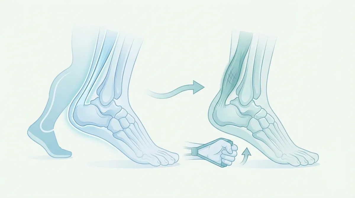
Title: Tendinopathies After 50: Why Tendons Suffer and How to Strengthen Them
The aging of the population and the parallel increase in active life expectancy have led to a significant increase in musculoskeletal pathologies. Among these, tendon affections represent one of the main causes of pain and disability in the adult population. Addressing an over 50 tendinopathy requires a deep understanding of the physiological, biomechanical, and structural changes that affect connective tissues with advancing age. Maintaining an active lifestyle is fundamental for cardiovascular and metabolic health, but it exposes tissues to mechanical loads that, if not adequately managed, can exceed the tendon’s own adaptive capacity. This article analyzes in detail the etiology, risk factors, most affected anatomical sites, and evidence-based strategies for the management and rehabilitation of tendinopathies in the sixth decade of life and beyond, always remembering that every therapeutic path must be supervised by a doctor or physical therapist.
- Tendinopathies are a significant cause of pain and disability in adults over 50.
- Aging leads to physiological and structural changes, increasing tendon vulnerability.
- Active lifestyles can expose aging tendons to loads beyond their adaptive capacity.
- Effective management requires understanding age-related changes and tailored rehabilitation strategies.
What is over 50 tendinopathy and why does it manifest
The term “tendinopathy” is now preferred by the international scientific community to describe a clinical picture characterized by pain, swelling (often absent in chronic phases), and functional limitation affecting a tendon. Historically, the terms “tendinitis” were used to indicate an acute inflammatory process and “tendinosis” to describe chronic degeneration without inflammation. However, modern research has shown that **over 50 tendinopathy** is a complex process, a true pathological “continuum” (Cook & Purdam, 2009) that includes reactive phases, dysrepair phases, and degenerative phases.
Anatomy and physiology of the aging tendon
Tendons are viscoelastic structures composed predominantly of water (about 60-80% of wet weight) and type I collagen (about 85% of dry weight), organized into parallel fibrils that confer high tensile strength. After the age of 50, the metabolism of tenocytes (the cells responsible for the synthesis and remodeling of the extracellular matrix) undergoes a physiological slowdown. There is a decrease in the synthesis of type I collagen in favor of type III collagen, which is less resistant and more disorganized (Magnan et al., 2014).
Furthermore, aging involves an increase in non-enzymatic cross-links (cross-linking) between collagen molecules, a process known as advanced glycation end-products (AGEs). This phenomenon makes the tendon stiffer and less able to dissipate elastic energy during movement, increasing susceptibility to micro-injuries. Vascularization, already naturally scarce in some critical areas (such as the mid-portion of the Achilles tendon or the supraspinatus insertion), further decreases, compromising the tissue’s reparative capacities.
The tendinopathy continuum model
According to the model proposed by Cook and Purdam, tendon pathology develops through three main stages, which often coexist in different portions of the same tendon, especially in subjects over 50:
- Reactive tendinopathy: An acute response to a sudden overload (e.g., a drastic increase in sports or work activity). The tendon thickens to reduce stress, but there is not yet true structural degeneration.
- Tendon Dysrepair: If the overload persists, the extracellular matrix begins to disorganize. There is an increase in proteoglycans that attract water, causing tissue swelling and separation of collagen fibers.
- Degenerative tendinopathy: Typical of advanced age and chronic pathologies. Cell death (apoptosis), acellular areas, disorganized neovascularization, and irreversible alteration of the matrix are observed. In this phase, the risk of spontaneous tendon rupture increases significantly.
Risk factors for over 50 tendinopathy
The onset of tendinopathy is almost never attributable to a single cause, but rather to a complex interaction between intrinsic factors (related to the individual) and extrinsic factors (related to the environment, mechanical load, and lifestyle). Identifying these factors is the first step that the doctor or physical therapist takes during the anamnesis.
Intrinsic factors: genetics, metabolism, and hormones
Age over 50 years itself represents a primary risk factor due to cellular senescence. Added to this are important metabolic alterations. Metabolic syndrome, obesity, and type 2 diabetes mellitus are strongly correlated with the development of tendinopathies. Chronic hyperglycemia accelerates the formation of advanced glycation end-products (AGEs), stiffening collagen. Central adiposity also generates a state of low-grade systemic inflammation through the release of adipokines, which alter tendon homeostasis (Leblanc et al., 2017).
In women, menopause plays a crucial role. The drastic drop in estrogen negatively affects collagen synthesis and the lubrication of connective tissues, explaining the high incidence of tendinopathies (particularly in the rotator cuff and gluteal tendons) in post-menopausal women.
Extrinsic factors: the role of biomechanical overload
The most common trigger remains an error in mechanical load management. This can translate into “overuse” (repeated overload over time) or a sudden peak load for which the tendon was unprepared. In amateur athletes over 50, the typical error is the attempt to maintain the training volumes and intensities of previous decades without allowing the body the necessary recovery times, which physiologically lengthen with age.
INAIL data and at-risk professional categories
Tendinopathies do not only affect athletes but represent one of the main causes of occupational disease. According to INAIL data on reported occupational diseases, musculoskeletal disorders account for over 65% of all reported pathologies. The over 50 age group is the most affected, due to the cumulative effect of repeated microtraumas over the working life.
Professional categories most exposed to biomechanical overload of the upper and lower limbs include:
| Professional Category | Biomechanical Risk Factors | Most Frequent Tendinopathies |
|---|---|---|
| Healthcare Workers (Nurses, OSS) | Manual handling of loads, incongruous postures, patient lifting. | Rotator cuff, epicondylitis, low back pain with myofascial involvement. |
| Construction and Building | Use of vibrating tools, heavy lifting, overhead work. | Rotator cuff, bicipital tendinopathy, epicondylitis. |
| Logistics and Warehousing | High-frequency repetitive movements, lifting loads from below. | Patellar tendinopathy, plantar fasciitis, epicondylitis. |
| VDU Workers | Prolonged static postures, repetitive mouse and keyboard use. | De Quervain’s tendinopathy, epicondylitis, epitrochleitis. |
Most affected anatomical sites after 50 years of age
Although any tendon in the human body can develop a pathology, daily biomechanics and vascular alterations make some sites particularly vulnerable in the over 50 population.
The Rotator Cuff (Shoulder)
The shoulder is the most mobile joint in the human body, but this mobility comes at a cost in terms of stability. The rotator cuff tendons (supraspinatus, infraspinatus, teres minor, and subscapularis) work incessantly to center the humeral head in the glenoid. After the age of 50, supraspinatus tendinopathy is extremely common. It is often associated with impingement syndrome (subacromial conflict), where the tendon is compressed between the humeral head and the acromion during arm elevation movements. Chronic degeneration in this area can lead to partial or full-thickness tears, often asymptomatic until an acute event triggers pain.
The Achilles Tendon
The Achilles tendon is the largest and strongest tendon in the body, fundamental for walking, running, and jumping. Achilles tendinopathy is divided into insertional (at the attachment to the calcaneus) and non-insertional (in the mid-portion, about 2-6 cm above the insertion). This latter area is known to be a watershed area of hypovascularization, which makes it particularly prone to degeneration in over 50 athletes (e.g., runners, tennis players). The classic symptom is morning stiffness that tends to improve with the first steps, only to flare up again after activity.
Epicondylitis and Epitrochleitis (Elbow)
Commonly known as “tennis elbow” (lateral epicondylitis) and “golfer’s elbow” (medial epitrochleitis), these pathologies affect the origin of the wrist and finger extensor and flexor muscles, respectively. Despite the names, most over 50 patients suffering from them do not play these sports, but develop the pathology due to repetitive work activities, prolonged computer use, or manual labor (e.g., gardening, DIY). Degeneration of the extensor carpi radialis brevis tendon is the most frequent anatomopathological finding in epicondylitis.
Gluteal Tendinopathy (Greater Trochanteric Pain Syndrome)
Often misdiagnosed as trochanteric bursitis, tendinopathy of the gluteus medius and minimus muscles is a primary cause of lateral hip pain, especially in women over 50. The pain typically manifests when lying on the affected side, climbing stairs, or walking uphill. Weakness of the hip abductor muscles and biomechanical alterations of the pelvis are the main contributing factors.
Plantar Fasciitis
Although not strictly a tendon, the plantar aponeurosis shares many histological and pathological characteristics with tendons. Plantar fasciitis is extremely common after 50 years of age, caused by the loss of elasticity of the plantar fat pad, alterations of the plantar arch (flat or high arch), and retractions of the posterior muscle chain (calf and hamstrings). Acute heel pain with the first steps in the morning is the pathognomonic symptom.
Symptomatology and clinical diagnosis
Timely recognition of symptoms is fundamental to prevent a reactive tendinopathy from turning into a chronic degenerative pathology. It is imperative not to rely on self-diagnosis, but always consult a doctor or physical therapist for an accurate clinical assessment.
Main symptoms
- Localized pain: The pain is usually pinpoint, well localized by the patient on the tendon or its bone insertion.
- “Warm-up” behavior: In the initial phases, pain is present at the beginning of activity, disappears or lessens during warm-up (a phase in which the tendon “warms up” and its compliance improves), only to reappear more acutely at rest, hours after exertion or the following morning.
- Morning stiffness: A sensation of tension and joint stiffness upon waking, which generally lasts from 10 to 30 minutes.
- Pain on palpation and loading: The tendon is painful when compressed, and symptoms are exacerbated when the corresponding muscle is required to generate force, especially during eccentric contraction (lengthening) or elastic energy storage (e.g., jumping).
The importance of diagnostic imaging
The diagnosis of tendinopathy is eminently clinical, based on anamnesis and provocative tests performed by the healthcare professional. However, instrumental imaging plays a crucial supporting role in excluding other pathologies (e.g., osteoarthritis, ligamentous injuries) and in evaluating the degree of structural degeneration (Docking et al., 2015).
- Musculoskeletal ultrasound: This is the first-line examination. It allows visualization of tendon thickening, the presence of hypoechoic areas (a sign of fiber disorganization and fluid accumulation), calcifications, and neovascularization (via Color or Power Doppler module).
- Magnetic Resonance Imaging (MRI): Used in more complex cases, when associated lesions are suspected (e.g., glenoid labrum tears in the shoulder) or when ultrasound is not conclusive. It provides excellent resolution of soft tissues and bone edema.
It is important to emphasize that alterations visible on instrumental examinations do not always correlate with pain. Many subjects over 50 present with degenerated tendons on ultrasound but are completely asymptomatic. Therefore, the instrumental examination must always be interpreted by the doctor or physical therapist in light of the patient’s clinical picture.
Conservative management and physiotherapeutic rehabilitation
The treatment of tendon pathology has undergone a revolution in the last two decades. The traditional approach based on absolute rest and non-steroidal anti-inflammatory drugs (NSAIDs) has been largely superseded. Today we know that “rest rusts” and that the tendon needs mechanical load to heal and remodel. Management must be multimodal and personalized.
Load Management
The first and most important therapeutic step is load modulation. This does not mean stopping completely, but identifying and temporarily reducing activities that cause excessive pain (greater than 3-4 on a scale of 0 to 10) or that cause a flare-up of symptoms within the next 24 hours. This process, known as “pacing,” allows the tendon to exit the reactive phase. For example, an over 50 runner with Achilles tendinopathy might be asked to temporarily stop uphill running or fast repeats, replacing them with swimming, cycling, or flat running at a gentle pace, thus maintaining aerobic capacity without overloading the tendon.
Therapeutic exercise: the heart of rehabilitation
Targeted physical exercise is the only intervention that has been shown to be able to modify the structure and load capacity of the tendon in the long term. The rehabilitation program, prescribed and progressed by the doctor or physical therapist, generally develops in several phases:
1. Isometric Exercises
In acute and highly reactive phases, isometric contractions (muscle contraction without joint movement) are the treatment of choice. Recent studies (Rio et al., 2015) have shown that heavy isometrics (e.g., 5 sets of 45 seconds with high loads) have a powerful analgesic effect, reducing cortical inhibition and alleviating tendon pain for several hours, allowing the rehabilitation process to begin safely.
2. Eccentric Training (Alfredson Protocol)
Eccentric exercise (the phase in which the muscle lengthens while developing tension) has been the gold standard for years, particularly for the Achilles tendon and patellar tendon. The protocol involves performing slow, controlled exercises during the lowering phase, often even in the presence of mild pain. Eccentric mechanical loading stimulates tenocytes to produce new collagen and helps realign disorganized fibers.
3. Heavy Slow Resistance (HSR)
More recent research (Kongsgaard et al., 2009) has introduced the concept of Heavy Slow Resistance training. This approach involves the use of high loads (70-85% of maximal) performed slowly in both the concentric (lifting) and eccentric (lowering) phases. HSR has proven to be as effective as pure eccentric training, but is often more agreeable to patients and produces better adaptations also at the level of the extracellular matrix and global muscle strength.
4. Energy Storage and Release Exercises (Plyometrics)
This final phase is essential for over 50 patients who wish to return to sports activities that require jumping, sprinting, or changes of direction (e.g., tennis, padel, running). It consists of gradually introducing fast and elastic loads (e.g., skipping rope, bounding) to re-accustom the tendon to its “spring” function.
Instrumental physical therapies and infiltrative therapies
Physical therapies can be a useful complement to therapeutic exercise, especially for pain control, but should never replace it.
- Extracorporeal Shockwave Therapy (ESWT): Particularly effective in insertional tendinopathies and in the presence of calcifications (e.g., calcific tendinopathy of the shoulder). High-energy acoustic waves stimulate neoangiogenesis (formation of new blood vessels) and tissue repair processes.
- High-Power Laser Therapy (HILT) and Tecar Therapy: Can promote the reduction of peritendinous edema and modulate pain in acute phases, improving local microcirculation.
As for infiltrative therapies, the use of corticosteroids is now strongly discouraged in chronic tendinopathies. Although they offer rapid short-term pain relief, in the long term they inhibit collagen synthesis, weaken the tendon structure, and significantly increase the risk of rupture. More modern alternatives, to be evaluated exclusively with the specialist doctor, include infiltrations of Hyaluronic Acid or PRP (Platelet-Rich Plasma), which aim to stimulate biological tissue regeneration.
Prevention and lifestyle for tendon health
Preventing an **over 50 tendinopathy** requires a holistic approach that goes beyond simple biomechanics, embracing lifestyle in its entirety.
Nutrition, hydration, and supplementation
The health of connective tissues strictly depends on nutrient intake. A diet rich in antioxidants (fruits, vegetables, omega-3s) helps counteract oxidative stress and systemic inflammation. Vitamin C is an essential cofactor for collagen synthesis; a deficiency compromises the stability of tendon fibers. Hydration is equally crucial: tendons need water to maintain their viscoelasticity and allow the sliding of tendon fascicles. The use of supplements based on hydrolyzed collagen peptides, associated with Vitamin C and taken approximately 60 minutes before physical exercise, is showing promising results in scientific literature for promoting extracellular matrix synthesis.
Warm-up, recovery, and flexibility
With advancing age, the time dedicated to warm-up must increase. A dynamic warm-up prepares tissues for load absorption, increasing local temperature and improving joint lubrication. Equally important is recovery (cool-down) and maintaining good muscle flexibility. Retracted or excessively stiff muscles transfer abnormal tensions to the tendons. However, prolonged static stretching before sports activity is discouraged, as it can temporarily reduce the tendon’s ability to store elastic energy; it is preferable to include it in dedicated sessions or at the end of training.
Finally, the choice of footwear (for lower limb pathologies) and sports equipment (e.g., racket string tension for epicondylitis) plays a fundamental preventive role. Workplace ergonomics, with active breaks and postural variations, is indispensable for preventing occupational overload pathologies highlighted by INAIL data.
Frequently Asked Questions (FAQ)
How long does it take to recover from tendinopathy after 50 years of age?
Recovery times vary considerably depending on the stage of the pathology and the affected site. Since tendon metabolism is slow, especially after 50 years of age, rehabilitation requires patience. A reactive tendinopathy can resolve in 3-6 weeks with adequate load management. However, a chronic degenerative tendinopathy requires a structured rehabilitation program that can last from 3 to 6 months, and sometimes up to a year, to achieve significant tissue remodeling. It is fundamental to follow the instructions of the doctor or physical therapist without rushing the process.
Can I continue training if I have tendon pain?
Yes, but with appropriate modifications. Absolute rest is counterproductive for tendons. The goal is “optimal load.” It is generally acceptable to continue training if the pain during activity remains mild (not exceeding 3-4 out of 10) and if there is no increase in stiffness or pain the following morning. If the pain worsens, it is necessary to reduce the volume, intensity, or frequency of training. The doctor or physical therapist will be able to indicate alternative activities (cross-training) to maintain physical fitness without overloading the injured tendon.
Are cortisone injections recommended for treating tendons?
Current international guidelines strongly advise against the use of corticosteroid injections for chronic tendinopathies. Although cortisone can provide rapid short-term pain relief due to its powerful anti-inflammatory effect, in the medium and long term it negatively alters the metabolism of tendon cells, inhibits collagen production, and significantly increases the risk of spontaneous tendon rupture. They should only be considered in exceptional cases and under strict medical evaluation.
Do weight exercises harm inflamed tendons?
On the contrary, exercise with resistance (weights) is the main therapy for tendinopathies. Tendons respond positively to heavy and slow mechanical load (Heavy Slow Resistance). The use of weights, if correctly dosed and included in a progressive program by the doctor or physical therapist, stimulates tenocytes to produce new collagen, increases the tensile strength of the tendon, and improves the strength of the associated muscle, reducing the risk of future recurrences.
Recommended Products for Rehabilitation Support
Product links are affiliate: purchasing does not incur additional costs for the user. These products do not replace the advice of a doctor or physical therapist.
Frequently Asked Questions
What is tendinopathy and why is it particularly relevant for individuals over 50?
Tendinopathy describes a clinical condition characterized by tendon pain, functional limitation, and sometimes swelling, reflecting a complex pathological continuum rather than simple inflammation. In individuals over 50, age-related physiological and structural changes increase tendon vulnerability, making them more susceptible to developing this condition.
How do age-related changes contribute to the development of tendinopathies after 50?
With advancing age, connective tissues undergo significant physiological, biomechanical, and structural changes that diminish a tendon’s adaptive capacity. These alterations can make tendons less resilient to mechanical loads, increasing the risk of developing pain and disability even with activities that were previously well-tolerated.
Can maintaining an active lifestyle increase the risk of tendinopathy in older adults?
While an active lifestyle is crucial for overall health, it can expose aging tendons to mechanical loads that may exceed their reduced adaptive capacity. Without adequate management and appropriate loading strategies, this exposure can contribute to the onset or exacerbation of tendinopathies in individuals over 50.
What are the general principles for managing tendinopathies in individuals over 50?
Effective management of tendinopathies in this age group requires a deep understanding of age-related tissue changes and tailored rehabilitation strategies. Therapeutic paths typically involve evidence-based approaches focused on strengthening and load management, always under the supervision of a doctor or physical therapist.
Sources and Scientific References
- Arya S, Kulig K. Aging and tendinopathy: a review of the cellular and molecular mechanisms. J Orthop Sports Phys Ther. 2010;40
Sources and Scientific References
- Breda SJ et al. (2021). Effectiveness of progressive tendon-loading exercise therapy in patients with patellar tendinopathy: a randomised clinical trial. Br J Sports Med. 55:501-509. DOI | PubMed
- Theodorou A et al. (2023). Patellar tendinopathy: an overview of prevalence, risk factors, screening, diagnosis, treatment and prevention. Arch Orthop Trauma Surg. 143:6695-6705. DOI | PubMed
- Beyer R et al. (2015). Heavy Slow Resistance Versus Eccentric Training as Treatment for Achilles Tendinopathy: A Randomized Controlled Trial. Am J Sports Med. 43:1704-11. DOI | PubMed
- Maffulli N et al. (2020). Achilles tendinopathy. Foot Ankle Surg. 26:240-249. DOI | PubMed
- Özmen T et al. (2021). Comparison of the clinical and sonographic effects of ultrasound therapy, extracorporeal shock wave therapy, and Kinesio taping in lateral epicondylitis. Turk J Med Sci. 51:76-83. DOI | PubMed